Home

Preferencinis gydymas jausti Neteisinga t1 t2 spine Glaustis kompensuoti Pati

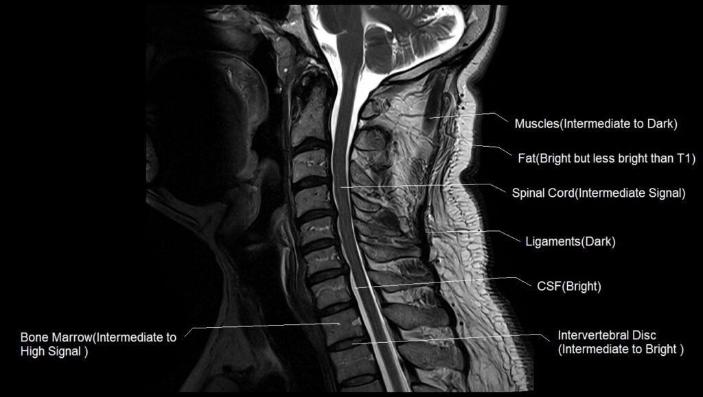
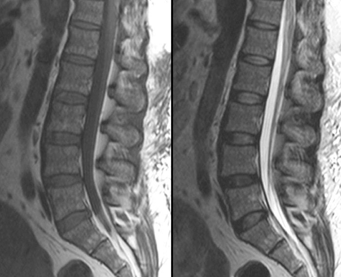
MRI interpretation - T1 v T2 images

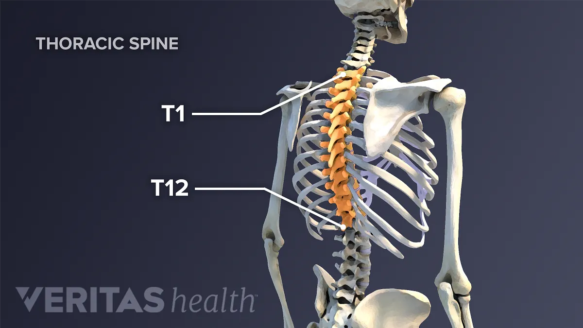
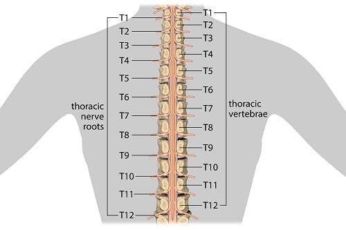
Thoracic Spinal Nerves | Spine-health

View Image
View Image

MRI whole spine, sagittal view. (A) T2 weighted and (B) T1 weighted... |  Download Scientific Diagram
MRI whole spine, sagittal view. (A) T2 weighted and (B) T1 weighted... | Download Scientific Diagram

MRI of Spinal Bone Marrow: Part 2, T1-Weighted Imaging-Based Differential  Diagnosis | AJR
MRI of Spinal Bone Marrow: Part 2, T1-Weighted Imaging-Based Differential Diagnosis | AJR

T1 vs T2 MRI | T1and T2 MRI image comparison

Thoracic Spinal Cord Injury: Functions Affected & Recovery

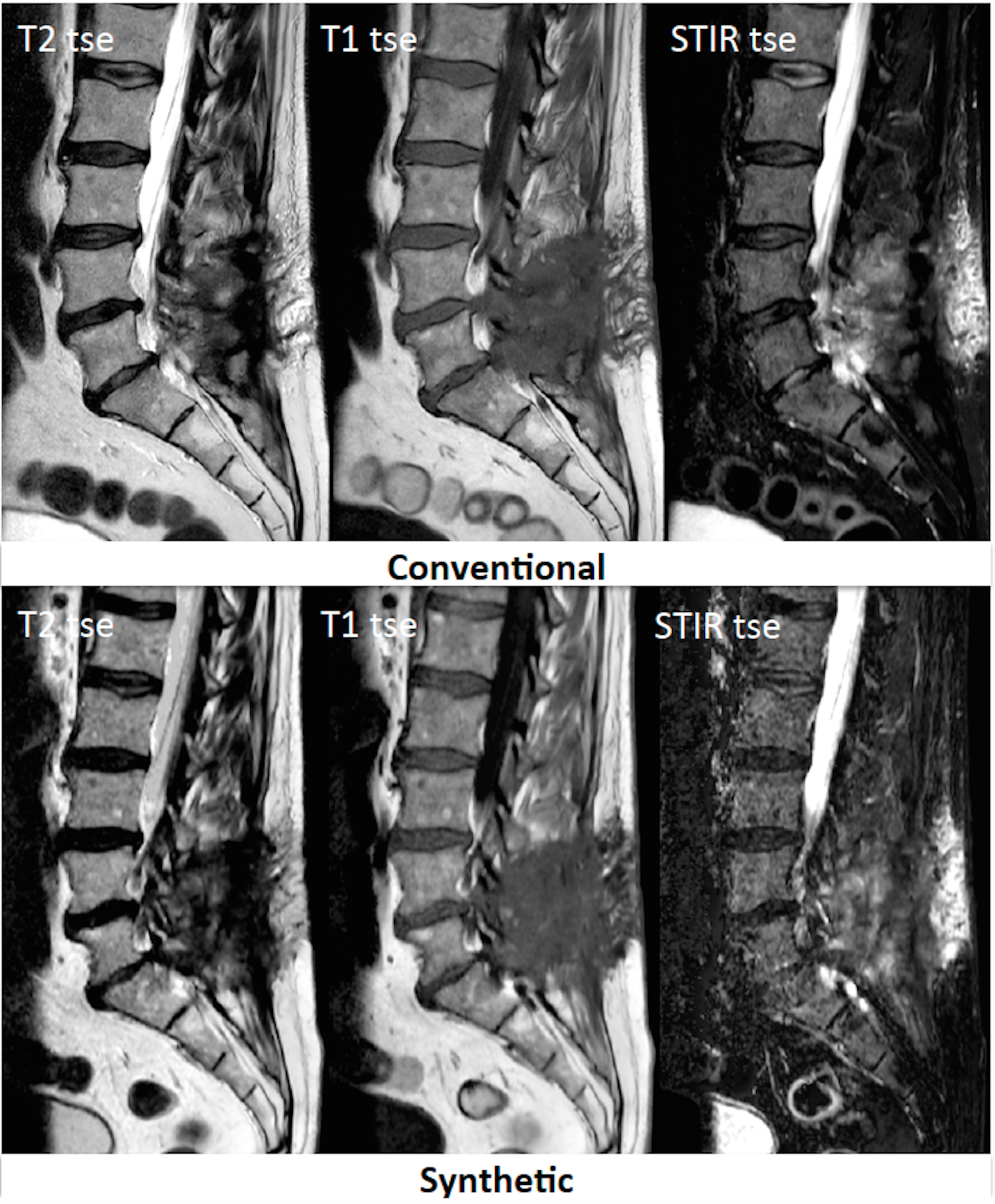
Comparing T1-weighted and T2-weighted three-point Dixon technique with  conventional T1-weighted fat-saturation and short-tau inversion recovery  (STIR) techniques for the study of the lumbar spine in a short-bore MRI  machine - ScienceDirect
Comparing T1-weighted and T2-weighted three-point Dixon technique with conventional T1-weighted fat-saturation and short-tau inversion recovery (STIR) techniques for the study of the lumbar spine in a short-bore MRI machine - ScienceDirect

Preoperative MRI of thoracic spine: T2 (A) and T1-weigh | Open-i

Comparison of Sagittal FSE T2, STIR, and T1-Weighted Phase-Sensitive  Inversion Recovery in the Detection of Spinal Cord Lesions in MS at 3T |  American Journal of Neuroradiology
Comparison of Sagittal FSE T2, STIR, and T1-Weighted Phase-Sensitive Inversion Recovery in the Detection of Spinal Cord Lesions in MS at 3T | American Journal of Neuroradiology
EPOS™
EPOS™

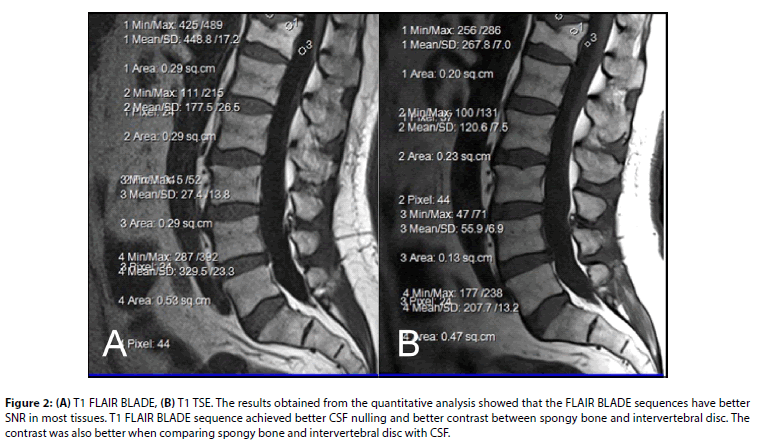
Comparison of T1 FLAIR BLADE with and without parallel imaging against T1  turbo spin echo in the MR imaging of lumbar spine in the sagittal plane
Comparison of T1 FLAIR BLADE with and without parallel imaging against T1 turbo spin echo in the MR imaging of lumbar spine in the sagittal plane

Diagnostics | Free Full-Text | Primary Benign Neoplasms of the Spine

Feasibility of Synthetic MRI of the spine – fast and quantitative imaging

A Rare Case of T1-2 Thoracic Disc Herniation Mimicking Cervical  Radiculopathy | International Journal of Spine Surgery
A Rare Case of T1-2 Thoracic Disc Herniation Mimicking Cervical Radiculopathy | International Journal of Spine Surgery

thoracic spine | Musculoskeletal Key

What do T1 and T2 weighted images mean in an MRI? - Quora

Neurological Examination Spinal Cord Part 3 - Everything You Need To Know -  Dr. Nabil Ebraheim - YouTube
Neurological Examination Spinal Cord Part 3 - Everything You Need To Know - Dr. Nabil Ebraheim - YouTube

T1 vs T2 MRI | T1and T2 MRI image comparison

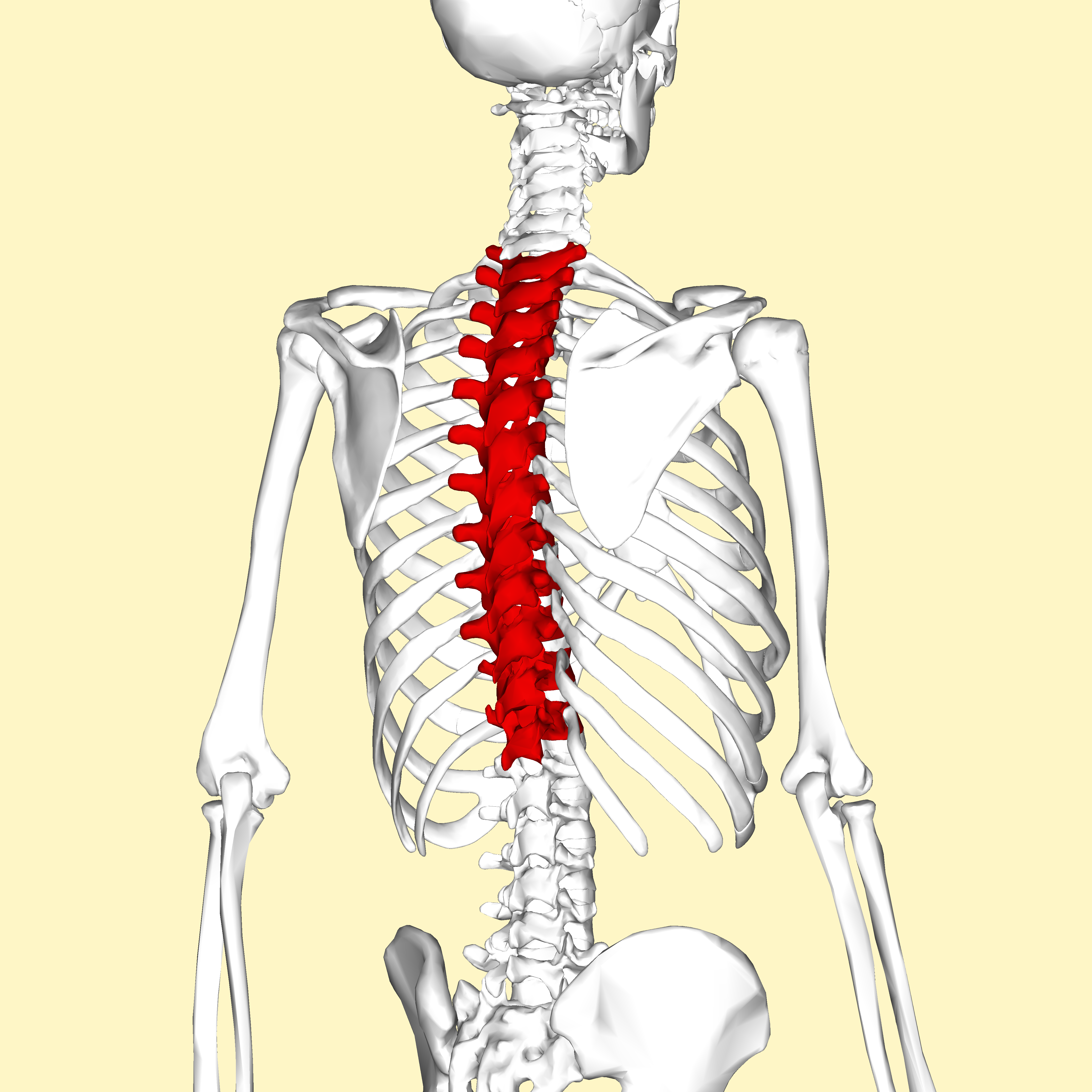
Thoracic vertebrae - Wikipedia

Intelligent Chiropractic - T1, T2, & T3: The Top Three Thoracic Vertebrae  Tension, Tightness, & Tech Neck Posture are also 3 T's associated with  symptoms that affect your upper thoracic spine. When
Intelligent Chiropractic - T1, T2, & T3: The Top Three Thoracic Vertebrae Tension, Tightness, & Tech Neck Posture are also 3 T's associated with symptoms that affect your upper thoracic spine. When
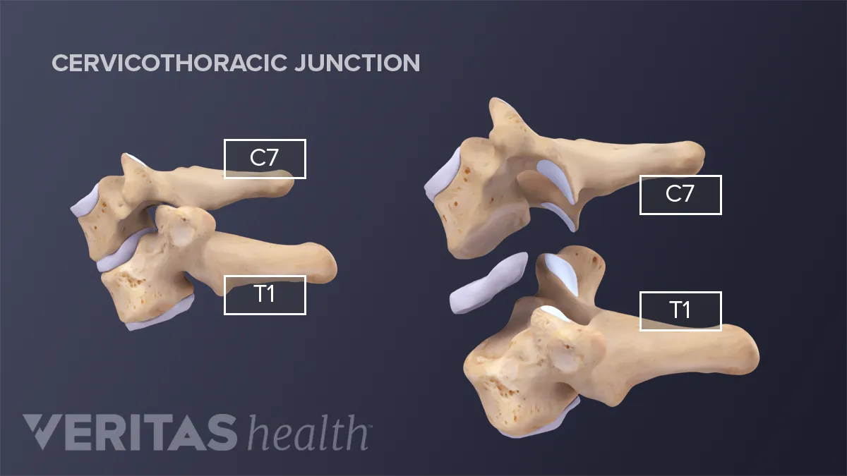
All About the C7-T1 Spinal Segment (Cervicothoracic Junction) | Spine-health

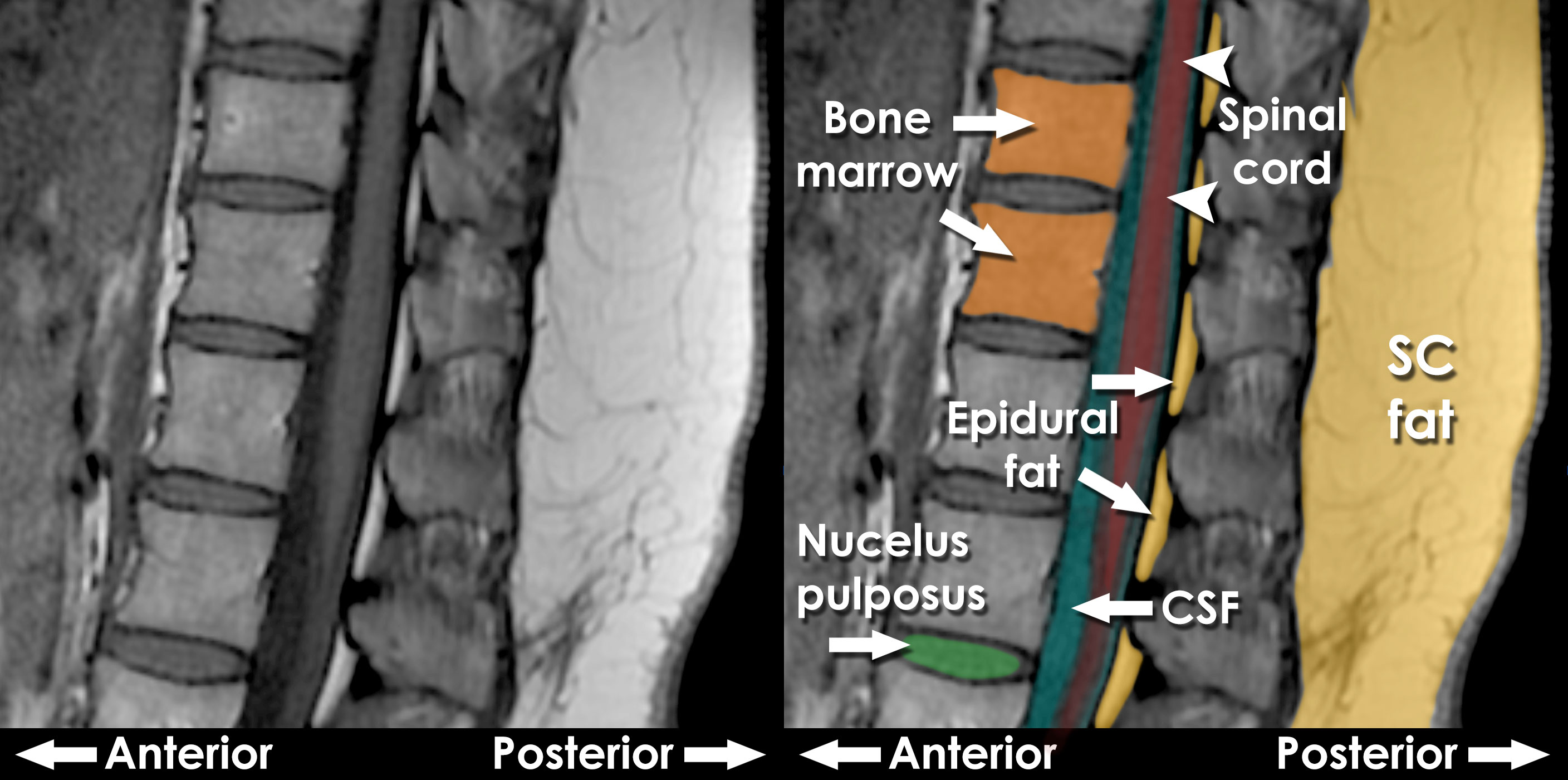
Normal Sagittal MRI
Normal Sagittal MRI

View Image
View Image

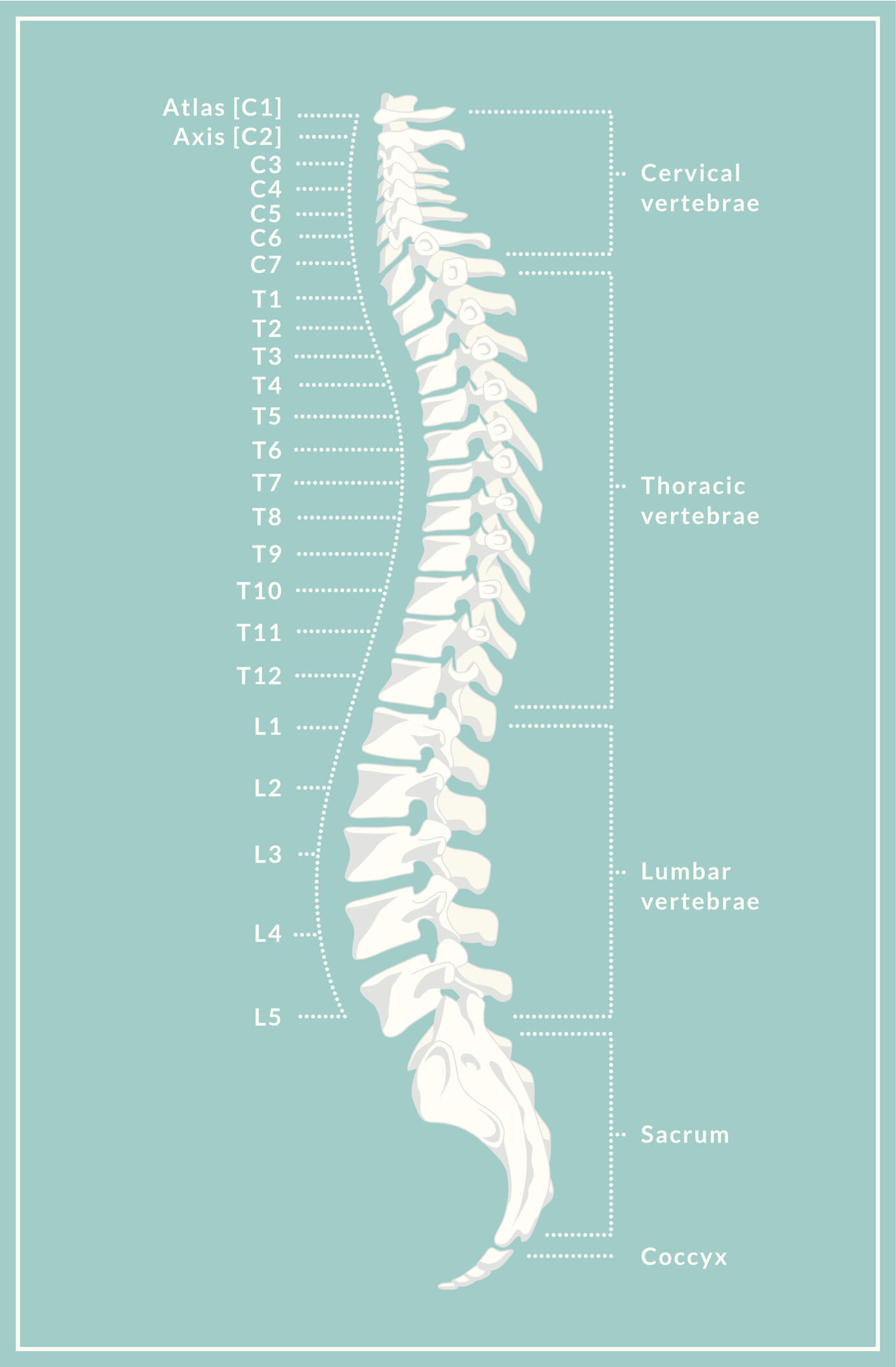
Spinal Anatomy Overview - Atlanta Brain and Spine Care

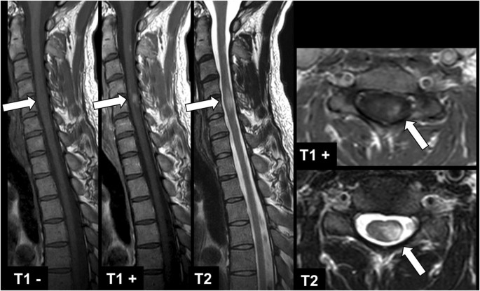
Pre-contrast T1-weighted imaging of the spinal cord may be unnecessary in  patients with multiple sclerosis | European Radiology
Pre-contrast T1-weighted imaging of the spinal cord may be unnecessary in patients with multiple sclerosis | European Radiology